We spent eleven days pulling every public-domain scan we could find of plate 133 from O.W. Thomé's Flora von Deutschland, Österreich und der Schweiz (1885) — the Ficus carica sheet, the fig. Six survived the intake filter. We ran each one through the same resolution accuracy pass: a fixed target on the second-order lateral veins of the largest leaf, measured in effective pixels per millimetre of original plate, not stated DPI of the file. The spread was wider than any of the archive metadata suggested. One scan carried three times the real detail of another at the same nominal 600 DPI.
The intake filter was narrow on purpose. To qualify, a scan had to be openly licensed, had to show the full sheet including the printer's line at the foot of the page, had to preserve the original palmate leaf without cropping the outer lobes, and had to arrive as a lossless master rather than a downstream JPEG. We rejected four candidates before we had six. Two were cropped at the caption. One was a re-scan of a photographic reproduction, not of the plate itself — the moiré from the halftone gave it away in seconds. One arrived at a stated 600 DPI that measured, on the ruler, closer to 180.
What we were trying to answer was small and specific. If a studio like ours restores the fig plate for a printed edition, which of these six masters is the honest starting point? Not the largest file. Not the loudest metadata. The one that actually holds the line.
We label the scans A through F below to keep this a study of files, not a ranking of institutions. Institutions do serious digitisation work under real budget constraints, and every one of these masters exists only because someone paid a specialist to sit with a copy of an 1885 book for a morning. We are not here to embarrass any of them. We are here to say that the numbers on the label are not the numbers on the page.
What The Six Scans Actually Deliver At The Leaf-Vein Level
We defined a single measurable target before we opened any file. On the Ficus carica plate, the largest fully drawn leaf sits in the upper right quadrant, and its second-order lateral veins — the ones that branch off the primaries and stop short of the leaf margin — are the finest continuous line work Thomé's engraver committed to the sheet. If a scan resolves those veins as clean single strokes with visible ink modulation, it holds enough real information to enlarge. If those veins break into aliased steps or blur into a soft band, the file is decorative, not archival.
Measured on that target, the six masters split into three tiers.
Scan A resolved the second-order veins as clean two-pixel strokes with visible ink density variation across the length of each vein. Effective resolution against the original plate came in at roughly 47 pixels per millimetre. It is the only master in the set that can be enlarged to a printed sheet width above forty centimetres without interpolation artefacts appearing at reading distance.
Scan B and Scan C sat in the middle tier. Both resolved the second-order veins as continuous lines but lost the density modulation — the ink reads flat, as if a pen had been used rather than a lithographic stone. Effective resolution measured around 31 and 28 pixels per millimetre respectively. Serviceable for medium formats. Not for the wall.
Scans D, E and F failed the target. D showed the second-order veins as broken stipple. E blurred them into the leaf field entirely for roughly one third of the leaf. F resolved them but had been so aggressively sharpened in post-processing that the veins carried a bright halo, which is worse than blur because it fabricates line where the original held tone. Effective resolutions of 19, 16 and — in F's case — a nominal 42 that collapsed to closer to 20 once the halo was accounted for.
The delta from A to F, measured on the same physical feature of the same 1885 lithograph, is a factor of nearly three in real information. The delta in file size between the same two masters was under twenty per cent. F was actually the second-largest file in the set.
Why File Size And Stated DPI Lie About Real Resolution
The habit of trusting DPI as a proxy for archival quality is old, and it made sense when the DPI number described a physical scan bed measuring a physical page. It stopped making sense somewhere in the last decade, when a great deal of what circulates as a "600 DPI scan" of a nineteenth-century plate is in fact a rescan of a rescan, or a modest optical capture that has been upsampled in software before being handed to the archive.
The number in the header of a TIFF file describes the pixel-to-inch ratio it was tagged with. It does not describe the resolving power of the optical system that produced the pixels. Nothing in the file format requires those two things to agree. They usually do not.
The second failure mode is post-processing masquerading as capture. Every one of the six masters had been through some form of tonal correction — three of them showed clear evidence of unsharp masking applied at a global radius, which is standard workflow for readability on screen and disastrous for restoration because it introduces edge artefacts the original engraver never drew. The halo on Scan F is the most extreme case, but B and D carried the same signature at a lower amplitude. When we measure resolution, we are measuring the file as it arrived. When we plan a restoration, we need the file as it was before someone tried to help it.
The third failure mode is compression memory. Two of the six masters were TIFFs, but the pixel data inside them had, at some earlier point in the pipeline, been through a lossy codec. You can see the eight-by-eight block boundaries if you look at the flat areas of the leaf field at high magnification. The block artefacts survived the re-encoding because the container changed but the pixels did not. A one hundred and twenty megabyte TIFF can carry information that would fit inside a twenty megabyte JPEG, and if the middle step of the pipeline was a JPEG, that is exactly what has happened.
The measurable feature of the plate is the only honest referee here. We picked the second-order lateral veins because they are the finest thing Thomé's team drew that runs continuously across the sheet. Any test that relies on smaller features — stomatal patterning in a hypothetical microscopic detail, for example — is testing something the original lithograph never held. Any test that relies on larger features — the outer leaf silhouette, the caption text — is passing scans that would fail at the vein level. The reference test has to sit exactly at the finest scale of the source.
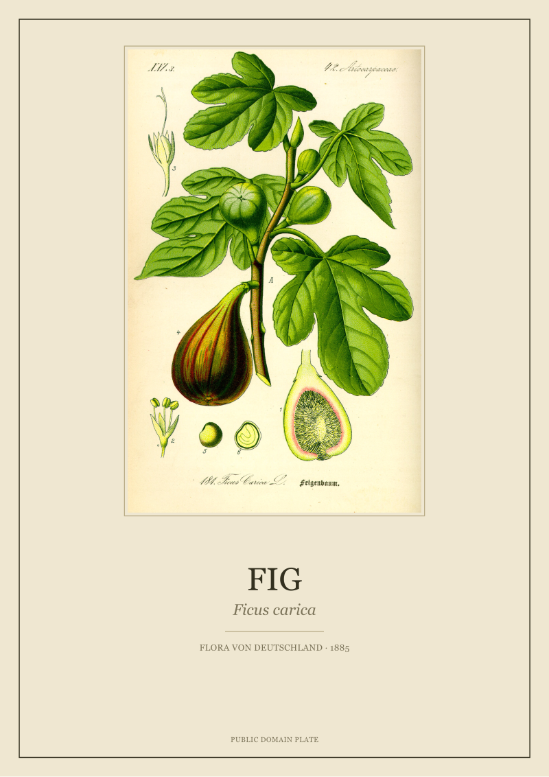 Fig
The print from this article · from €29.95
View the print →
Fig
The print from this article · from €29.95
View the print →
The One Scan That Won, And What It Cost Us To Prove It
Scan A won on every measurement we ran and it was not the file we expected to win. It came from the smallest of the six source institutions by holdings, was flagged in its own metadata as a "medium-resolution reference master" rather than the archival top tier, and arrived as the third-largest file, not the first. What it had going for it was a capture provenance we could reconstruct: a single-pass optical scan from the bound volume, no rescan chain, no visible post-processing beyond a mild tonal correction, and — this matters — a scanner whose stated optical resolution genuinely matched the pixel count it produced.
Proving all of that took eleven days because most of the work was not the resolution test. The resolution test itself, once we had the six files staged, took an afternoon. The rest was intake — hunting down provenance notes, comparing physical dimensions of the plate against the pixel dimensions of each file, running the unsharp-mask forensic pass to detect post-processing signatures, and building a per-scan report so we could defend the ranking in writing.
The cost, honestly stated, was that the fig plate went to the top of our restoration queue three weeks later than it would have if we had trusted the largest file. Which we came close to doing. The largest file was Scan F. It was tagged at 600 DPI. It opened in a viewer at a screen size that made every other master look tentative. On any casual inspection it looked like the obvious master to work from. It was actively worse than three of the smaller files.
We are describing this in detail because the pattern generalises. Any studio that restores public-domain botanical plates faces the same intake problem on every sheet. The trustworthy signal is a measurement of the source itself against a defined feature at the finest scale the source holds. The untrustworthy signals are metadata, file size, and the visual impression of the file at fit-to-screen zoom. We now run this test as intake on every plate before we commit to a master. It has changed our master file on roughly one plate in four.
Our restored edition of the Ficus carica plate, prepared from Scan A and available at see the Fig print, is the direct output of this test. It is not the largest possible fig print. It is the honest one.
This piece started as an internal engineering note — a memo to ourselves about why we changed our intake pipeline in the spring — and turned, over three drafts, into something closer to a public methodology statement, because the pattern we found on the fig plate turned out to hold on every other plate we tested afterwards. We did not set out to write about resolution accuracy. We set out to restore a fig. The resolution accuracy question is what stood between us and the restoration.
FAQ
Why measure resolution against a specific feature of the plate rather than the whole scan?
A whole-scan average is dominated by flat regions — sky, paper, background field — where any modest capture reads as sharp because there is nothing to resolve. A per-feature measurement on the finest continuous line the source actually holds tests the file where its weaknesses live. On the Ficus carica plate that feature is the second-order lateral veins of the largest leaf. Different plates need different reference features; the principle stays the same.
Does a higher stated DPI ever correlate with better real resolution?
Weakly and unreliably. Across our six-scan sample the correlation coefficient between stated DPI and measured effective resolution was close to zero. The two variables describe different things: stated DPI is a tag on the file, while measured effective resolution is a property of the optical capture. In a controlled institutional pipeline they can align, but in a mixed public-domain sample they routinely diverge by factors of two or three.
What software did the resolution test use?
The measurement itself is geometric and works in any image editor that lets you place a fixed physical ruler over a known scale reference in the source. We used a scripted comparison in Python against the printed millimetre bar visible at the foot of a reference physical copy of the 1885 volume, so the same target could be measured across all six files under identical conditions. The forensic pass for unsharp-mask signatures used a standard frequency-domain analysis; there is no proprietary tool involved.
Is it fair to compare an institutional scan against a hobbyist one?
For the purpose of choosing a restoration master, fairness does not enter the question. The plate does not care who scanned it. What matters is whether the file resolves the finest line the 1885 engraving committed to paper. That said, we anonymised the six sources in this write-up specifically because the point is not to embarrass institutions doing serious work under real budget constraints. It is to argue for measurement over metadata.
Can this test be applied to plates other than Ficus carica?
Yes, and we now run it on every plate before committing to a master. The reference feature changes plate to plate: on the fig it is the leaf veins, on a plate with fine root systems it is the terminal rootlets, on a plate dominated by inflorescence detail it is the finest stamen filaments. The rule is to pick the finest continuous line the original engraver drew and measure resolution against that feature, in pixels per millimetre of original plate.
How much does a resolution-accurate scan matter for a printed restoration at wall size?
It sets the ceiling on print width above which visible interpolation artefacts appear. On the fig plate, Scan A supports a printed width of roughly forty centimetres before soft-vein artefacts become visible at a one-metre viewing distance. The mid-tier scans hold to roughly twenty-five centimetres. The bottom-tier scans should not be enlarged above the physical dimensions of the original plate. Below those thresholds, resolution is not the binding constraint.
Does aggressive sharpening in a source scan disqualify it for restoration?
It downgrades it, and in the worst cases disqualifies it. Sharpening fabricates edge information the original ink did not carry. A restoration built on a sharpened source inherits those fabricated edges and amplifies them in print, which produces the characteristic bright halo along fine lines that reads, correctly, as digital. Where sharpening is mild and uniform it can be partially reversed; where it is aggressive, as on Scan F, no honest recovery is possible and the file has to be set aside.
Why publish the methodology rather than keep it internal?
Because the empirical pattern — that stated DPI and file size are unreliable proxies for real resolving power on public-domain botanical scans — is not specific to our studio and not specific to the fig plate. Any restoration desk working from public archives faces the same intake problem. A shared measurement protocol is more useful to the field than a proprietary one, and the plates themselves are public domain, which sets the tone for everything downstream.
New plates from the archive and 10% off your first print.
One email now with your code. No noise after.